What are the clinical benefits of 3D vs 2D endoscopy? This question matters as hospitals assess safer, clearer, and more precise visual systems. Three-dimensional endoscopy can provide depth perception, helping clinicians judge tissue distance during delicate procedures. A small vessel may appear more distinct. A narrow anatomical space may feel less visually confusing.
Professor Jacques Marescaux, a pioneer in minimally invasive surgery, has said, “The goal of technology is to improve patient care.” That principle remains important when comparing 3D and 2D endoscopy. 3D imaging may improve hand-eye coordination, instrument positioning, and spatial orientation. These advantages can support complex endoscopic surgery, especially when surgeons must dissect, clip, or suture within confined spaces.
The benefits are not automatic. Some clinicians experience eye strain, dizziness, or slower adaptation with 3D systems. Training quality, equipment calibration, operative complexity, and surgeon experience also influence outcomes. In routine examinations, 2D endoscopy may remain practical, familiar, and cost-effective. That is the less exciting answer.
Clinical evidence is still developing. Some studies report shorter task times and improved precision with 3D visualization, while others find limited differences in real-world patient outcomes. Therefore, hospitals should assess ergonomics, learning curves, maintenance costs, and procedure-specific results. The strongest choice is not always the newest system. It is the system that helps trained professionals make safer decisions, consistently.
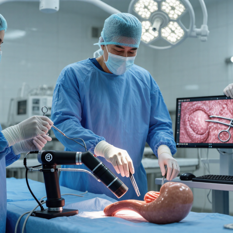
Three-dimensional (3D) and two-dimensional (2D) endoscopy use a flexible or rigid camera to display internal anatomy on a monitor. Both systems rely on illumination, image capture, and real-time visualization. The main difference is depth perception. A 2D image shows height and width, while a 3D system combines slightly different views to create spatial depth.
In clinical practice, 2D endoscopy remains familiar and widely used. It can provide clear images with simpler viewing requirements. 3D endoscopy may help clinicians judge distance, tissue layers, and instrument movement during selected procedures. Depth can make delicate manipulation feel more natural. However, 3D images may require compatible displays, careful calibration, and user adaptation. Better depth does not automatically mean better clinical decisions. Anatomy, lighting, image resolution, and operator training still matter. Some users may also experience eye strain or discomfort, especially during long procedures.
Tips: Match the system to the procedure, not the marketing claim. Check image clarity, depth accuracy, and ergonomic comfort in realistic conditions. Training should include simulated tissue handling and instrument positioning. Small viewing errors can become important near fragile structures. Not every case needs depth. Review clinical evidence, staff feedback, and maintenance requirements before choosing an endoscopy platform. Reassessment is useful because an impressive first demonstration may not reflect routine performance.
China Best 3D vs 2D Endoscopy Clinical Benefits?
Three-dimensional endoscopy creates separate images for each eye. This can strengthen depth perception during delicate dissection. Surgeons may judge tissue planes more confidently around vessels, ducts, and narrow pelvic spaces. In a crowded operative field, a suture can appear closer to the instrument tip. That small visual cue may reduce unnecessary movements. Two-dimensional endoscopy remains clinically useful. It offers a familiar flat image with clear spatial relationships learned through experience. Some surgeons also find it less tiring during long procedures.
Image quality depends on more than dimensional viewing. Resolution, color accuracy, light distribution, image stability, and monitor performance all affect clinical judgment. A high-quality 2D image can outperform a poorly calibrated 3D system. Glare and motion may weaken the practical benefit of stereoscopic vision. The difference is not absolute. Patient anatomy and procedure complexity matter greatly.
Clinical studies often report improved task accuracy or shorter learning curves with 3D systems. However, findings vary across specialties, operators, and outcome measures. In real operating rooms, setup time, head position, visual adaptation, and surgeon fatigue deserve attention. I have found that depth perception feels valuable during precise needle placement, yet it does not replace anatomical knowledge. Human judgment remains imperfect. Careful training, stable imaging, and objective patient outcomes should guide the choice between 3D and 2D endoscopy.
Three-dimensional endoscopy can improve depth perception during minimally invasive procedures. It presents anatomy with visual separation, rather than a flat image. This may help surgeons judge tissue planes, vessel distance, and instrument position more accurately. The benefit is especially noticeable when working in narrow spaces. Pelvic, thoracic, urological, and gastrointestinal procedures may gain from this added spatial information.
Small details matter. A suture can appear easier to place near delicate tissue. A clip may be positioned with greater confidence. During dissection, depth cues can support steadier hand-eye coordination. Clinical evaluations often report shorter learning curves for selected tasks, although results vary between procedures and operators. Experience still matters. A high-resolution 3D image cannot replace anatomical knowledge, careful planning, or team communication.
There are limitations. Some users experience eye strain, dizziness, or discomfort during longer cases. Equipment setup may also require training and thoughtful operating-room workflow. Evidence is not equally strong across every specialty. In my view, 3D endoscopy should be assessed by patient outcomes, procedure time, error rates, and user comfort, not visual appeal alone. Two-dimensional systems remain practical and effective in many settings. More comparative research is needed, particularly for complex cases and long-term recovery.
Clinical Applications and Advantages of 2D Endoscopy
2D endoscopy remains a practical tool across many clinical settings. It supports examinations in gastroenterology, bronchoscopy, urology, and ear, nose, and throat care. The image appears on a flat monitor, yet it can show mucosal color, bleeding, inflammation, and suspicious tissue clearly. Many clinicians value its familiar controls and stable workflow. It also integrates well with existing recording and documentation systems. These advantages can reduce training demands and support efficient daily procedures.
Tips: Adjust white balance before examination. Keep the lens clean. Use gentle movement and controlled insufflation. Compare findings with previous images when available. Do not judge depth from one view alone.
2D endoscopy is often more accessible than advanced three-dimensional systems. Hospitals may benefit from lower equipment costs and simpler maintenance. It can be useful where procedure volume is high and technical resources vary. However, depth perception is limited. Overlapping anatomy may become difficult to interpret, especially during delicate dissection or navigation. Image quality also depends on lighting, focus, patient movement, and operator technique. Better equipment cannot replace careful training. Clinical decisions should combine endoscopic findings, patient history, imaging, and pathology when required. The method is effective, but not flawless. That limitation deserves honest discussion during system selection.
| Clinical Dimension | 2D Endoscopy | 3D Endoscopy | Clinical Interpretation |
|---|---|---|---|
| Image perception | Provides a two-dimensional image using conventional monocular visualization. | Provides binocular stereoscopic visualization that can improve perception of depth and spatial relationships. | The practical benefit of 3D is most relevant when depth judgment is important; 2D remains adequate for many routine examinations. |
| Routine diagnostic endoscopy | Widely used for inspection, documentation, lesion localization, surveillance, and follow-up examinations. | May provide additional spatial information, but is not required for most standard diagnostic examinations. | 2D offers a mature, familiar, and broadly applicable workflow for everyday diagnostic practice. |
| Biopsy and tissue sampling | Supports forceps positioning and sampling under direct visual guidance. | Depth cues may assist with instrument positioning in selected procedures. | For routine biopsy, operator experience, lesion access, and instrument control generally remain more important than image dimensionality. |
| Therapeutic procedures | Used for polypectomy, hemostasis, dilation, foreign-body removal, and other endoscopic interventions. | Can support spatial orientation during technically demanding maneuvers, suturing, dissection, or work in confined spaces. | 3D may be advantageous for selected advanced interventions, but clinical benefit depends on the procedure and operator training. |
| Laparoscopic and thoracoscopic surgery | Provides reliable visualization for many established minimally invasive procedures. | May improve depth perception during dissection, suturing, knot tying, and identification of tissue planes. | 3D visualization can be particularly useful for precision tasks, while 2D remains effective across a broad range of operations. |
| Endoscopic mucosal and submucosal dissection | Supports lesion marking, injection, incision, dissection, and hemostasis using established techniques. | Additional depth information may help with orientation around the submucosal layer and vessels. | 3D can be a helpful adjunct, but procedure quality still depends heavily on training, lesion characteristics, and hemostatic capability. |
| Learning curve | Familiar to most clinicians and compatible with conventional endoscopic training and video systems. | May shorten the time needed to understand spatial relationships for some psychomotor tasks, although users require adaptation to stereoscopic viewing. | Training outcomes vary by procedure, learner experience, display quality, and study design. |
| Depth perception limitations | Depth must be inferred from motion, shadows, focus, tissue deformation, and instrument movement. | Stereoscopic images provide a direct binocular depth cue, although visual fatigue or discomfort may occur in some users. | 3D addresses a recognized limitation of 2D imaging, but it does not eliminate the need for anatomical knowledge and procedural skill. |
| Image quality and visualization | Current high-definition 2D systems can provide detailed mucosal and tissue visualization. | Image quality depends on stereoscopic resolution, camera alignment, display technology, and the viewing conditions. | Dimensionality and image resolution are separate characteristics; a 3D image is not automatically clearer than a high-definition 2D image. |
| Ergonomics and user comfort | Generally familiar and does not require stereoscopic glasses or binocular viewing equipment. | Some systems require special displays or glasses; prolonged use may cause eye strain, headache, or visual discomfort in susceptible users. | 2D may be preferable when long procedures, staff comfort, or compatibility with existing displays is a priority. |
| Operating-room integration | Usually integrates easily with established recording, teaching, documentation, and display workflows. | May require compatible cameras, processors, monitors, recording systems, and staff training. | 2D often has lower implementation complexity, while 3D may require additional infrastructure and workflow planning. |
| Cost and accessibility | Typically more accessible because conventional equipment, accessories, and training pathways are widely available. | Additional imaging and display components may increase acquisition, maintenance, and training requirements. | 2D may offer a favorable value proposition where routine diagnostic and therapeutic needs are the main priority. |
| Evidence and outcome assessment | Has extensive clinical use and established outcomes across diagnostic and therapeutic endoscopy. | Studies often report improved depth perception or task performance, but consistent improvements in major patient outcomes are procedure-specific and not universal. | Equipment selection should be based on the intended procedure, clinical evidence, workflow, training, and total cost rather than visualization format alone. |
Note: Clinical performance varies with anatomy, procedure complexity, image resolution, equipment configuration, operator experience, and institutional workflow. 3D visualization should be considered an adjunct rather than a universal replacement for 2D endoscopy.
China Best 3D vs 2D Endoscopy Clinical Benefits?
Safety depends on more than image depth. A Cochrane review found that 3D systems often improve laboratory task accuracy and shorten task time. However, evidence for fewer complications remains limited and low-certainty. Better screen detail does not automatically mean safer surgery. Published trials frequently measure speed, not long-term recovery. That gap matters. Some operators experience eye strain, dizziness, or slower orientation during early use. Hospitals should monitor these effects through supervised cases and incident reviews.
Training requirements can change the financial equation. A 2022 meta-analysis in Surgical Endoscopy reported improved depth perception and hand-eye coordination with 3D visualization, especially during complex simulated tasks. Yet transfer to routine clinical outcomes was inconsistent. Training may require simulation time, mentor coverage, and structured competency checks. Equipment costs also include maintenance, compatible instruments, room integration, and staff education. A cheaper purchase can become expensive later.
Selection should match case complexity, surgeon experience, and operating-room capacity. High-volume units may value 3D during suturing, dissection, and narrow pelvic work. Lower-volume units may prefer dependable 2D systems with familiar workflows. Procurement teams should compare procedure time, error rates, training hours, and total ownership costs. The evidence is useful, but not perfect. Local audits may reveal a different answer.
It provides separate images for each eye. Tissue planes may appear clearer near vessels, ducts, and narrow pelvic spaces.
No. Resolution, color, lighting, stability, and monitor quality also matter. A well-calibrated 2D system may look better.
It may help with precise needle placement, suturing, and delicate dissection. A suture can appear closer to the instrument tip.
Yes. It offers a familiar flat image and dependable spatial relationships. Some surgeons experience less fatigue during long procedures.
Not automatically. Studies often show better task accuracy, but evidence for fewer complications remains limited.
Some users report eye strain, dizziness, or slower orientation. Supervised practice can reveal these problems early.
Costs include equipment, maintenance, compatible instruments, room integration, and staff education. A cheaper purchase may cost more later.
Consider case complexity, surgeon experience, training capacity, and room workflow. Local audits may provide a better answer than general claims.
No. Anatomical knowledge and careful judgment remain essential. I may overvalue visual detail without checking patient outcomes.
Track procedure time, errors, training hours, fatigue, and recovery outcomes. Speed alone is not enough.
What are the clinical benefits of 3D vs 2D endoscopy? This article compares the basic principles of both technologies and explains how they differ in visualization, depth perception, spatial orientation, and image quality. By presenting a more realistic sense of depth, 3D endoscopy may support improved hand-eye coordination, more accurate tissue manipulation, and greater confidence during procedures that require precise movements. These advantages can be valuable in minimally invasive surgery, training, and complex anatomical examinations.
2D endoscopy remains a practical and widely used option, offering clear images, familiar operation, broad compatibility, and generally lower equipment and training costs. The article also considers safety, operator learning curves, procedure type, patient needs, maintenance, and clinical workflow. Rather than identifying one system as universally superior, it emphasizes that the appropriate choice depends on the required level of spatial detail, available resources, staff expertise, and the specific medical application.

Centuri Medical