Circular staplers can simplify colorectal anastomosis, but they demand disciplined handling. A small technical mistake may crush tissue, create bleeding, or compromise the staple line. The risks often appear during ordinary moments: forceful insertion, poor visualization, uneven tissue capture, or hurried firing.
So, how can surgeons answer the question, “How to avoid accidental tissue trauma with circular staplers?” The solution is not one isolated maneuver. It involves careful patient selection, complete tissue alignment, gentle instrument passage, and deliberate confirmation before firing. Experienced teams also inspect the donuts, examine the staple line, and consider an air-leak test when clinically appropriate. These steps are practical, but they are not foolproof.
Colorectal surgeon Dr. Steven D. Wexner has emphasized a principle relevant to stapling safety: “Good surgery depends on precision, judgment, and respect for tissue.” That message deserves attention. A stapler should never be treated as a substitute for anatomy, visualization, or patience.
The following seven tips focus on preventable sources of trauma. They address tissue handling, device positioning, firing technique, and post-firing inspection. Some recommendations may seem obvious. They are still missed in real operating rooms.
A useful reminder: tissue rarely “complains” before damage occurs. Resistance, abnormal tension, or an incomplete tissue ring should prompt reassessment, not additional force. No checklist removes human error completely. However, a structured approach can expose weak points before they become complications.
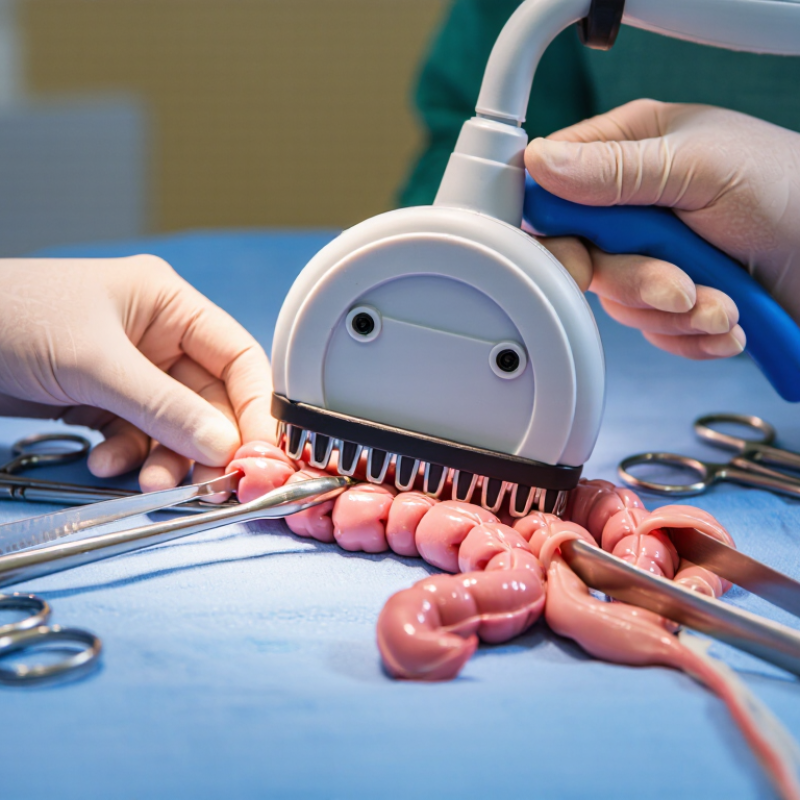
Tissue trauma during circular stapler use often begins before the device is fired. Edema, bruising, tension, or poor blood supply can make tissue less tolerant of compression. A careful assessment of tissue quality matters more than speed. The surgeon should confirm that the bowel reaches the anastomotic site without force. Excessive traction can create hidden tears.
Small details influence the injury pattern. The purse-string should capture an even tissue ring without excessive bites or strangulation. Tissue should lie flat around the shaft, without folds, twisting, or trapped mesentery. Gentle approximation allows compression to occur gradually. It may feel slower, but haste can damage the tissue edge. The stapler should be aligned with the bowel rather than angled against it. Firing should follow the patient’s anatomy and the device’s instructions, not routine alone.
After firing, inspect the anastomosis carefully. Both tissue rings should be complete, but complete rings do not prove perfect healing. Check for bleeding, gaps, tension, and abnormal discoloration. A leak test can provide useful evidence, although it cannot replace direct clinical judgment. In practice, surgeons may underestimate trauma when the tissue looks acceptable externally. That is worth questioning. Patient factors, tissue thickness, prior treatment, and local perfusion can change the result. Follow institutional protocols, document unexpected findings, and involve experienced colleagues when the anatomy or tissue quality is uncertain.
Selecting the correct circular stapler size begins with anatomy, not convenience. The device diameter should match the lumen without forcing tissue into the shaft. A stapler that is too large may stretch the bowel. One that is too small can create crowding, uneven compression, or a narrow anastomosis. Measure carefully, and avoid guessing from visual estimates.
Tissue thickness matters just as much. Thick, edematous, or poorly perfused tissue may exceed the instrument’s recommended compression range. Always compare the measured tissue with the stapler’s validated thickness information. The closed staple height must accommodate the tissue without crushing it excessively. A 2019 FDA safety communication recorded more than 41,000 reports involving surgical staplers and staples, including 366 deaths and over 9,000 serious injuries. These figures cover many stapler types, not circular devices alone, but they show why sizing cannot be casual.
During placement, keep the tissue flat and free from unintended folds. Check that the anvil is fully seated before firing. Do not pull aggressively to make the tissue fit. That shortcut can hide a poor match. After firing, inspect the tissue rings for completeness and examine the staple line for bleeding or gaps. Evidence from colorectal studies commonly links technical factors, including tissue thickness and incomplete doughnuts, with anastomotic complications. The difficult part is that real tissue rarely behaves like a measurement chart. Reassess when swelling, tension, or perfusion changes.
Before firing a circular stapler, tissue preparation deserves deliberate attention. Published colorectal studies and ACS NSQIP analyses report clinically important anastomotic leaks from low single digits to nearly 10%, depending on patient risk and procedure type. Poor alignment is not the only cause, but it can increase compression, bleeding, and incomplete staple formation.
Tips: Clear excess mesenteric fat from the intended staple line. Keep the bowel edges moist, evenly sized, and free from twisting. Check that the mesentery lies naturally, not trapped beneath the anvil or shaft. A gentle alignment check matters more than speed. The tissue should meet without tension, gaps, or forceful traction. Experienced teams also assess perfusion before firing, using visual color, pulsation, and established intraoperative assessment methods when available.
Do not ignore small folds.
Professional guidance from the ERAS Society and colorectal surgical standards supports careful handling, tension reduction, and perfusion assessment, although the exact technique depends on anatomy and operative judgment. After closing the stapler, confirm that tissue has not slipped or rotated. The doughnuts should be complete, but they are not a perfect safety guarantee. A technically complete ring can still accompany poor perfusion or excessive tension. This is where practice sometimes falls short: alignment may look acceptable from one angle, yet reveal a hidden fold from another. Pause, rotate the field, and reassess before firing.
Circular staplers can reduce operative time, but tissue trauma often begins before firing. The key is controlled compression, not maximum compression.
Bring the tissue together evenly, without pulling one side tighter than the other. Uneven capture can create a thick, bruised edge and a weak staple line. Inspect the tissue before closing. Fat, folds, or trapped mesentery may distort the compression zone.
A short pause after closing can help tissue settle. Follow the device instructions and local surgical protocol for the recommended compression time. Do not rush the trigger.
During firing, keep the instrument stable and avoid twisting the shaft. A smooth, continuous motion is usually safer than repeated hesitation. Excessive force can crush tissue, while insufficient closure may leave gaps. Both problems can be difficult to detect immediately.
After firing, examine the staple line carefully. Look for bleeding, incomplete tissue rings, uneven staple formation, or a visibly thin segment. Leak testing should match the procedure and institutional practice.
Small details matter here. I have seen good preparation weakened by a hurried final squeeze. That mistake is easy to understand, but it deserves honest review.
Team communication also helps: the assistant should confirm tissue position, alignment, and any unexpected resistance before firing.
Inspecting the anastomosis begins before removing the protective instruments. Look for even staple formation, aligned edges, active bleeding, and tissue caught within the closure. The surrounding bowel should appear warm, moist, and reasonably perfused. A small ooze matters. Gentle suction can reveal bleeding that looks harmless at first. Avoid pulling the anastomosis to improve visibility, because traction may enlarge a minor injury.
A controlled leak test may help, when appropriate for the procedure and local protocol. Observe the entire circumference, not only the easiest side to reach. Check for bubbles, separation, narrowing, or a folded edge. If the tissue appears bruised, pale, or torn, pause and reassess its viability. The safest response depends on the injury, blood supply, tension, and the patient’s condition. A quick reinforcement is not always a complete solution.
After surgery, early tissue injury may present through increasing pain, fever, rapid heart rate, abdominal distension, or unexpected drainage. These signs can be subtle. Do not rely on one reassuring observation. Serial examinations, laboratory trends, and imaging should support clinical judgment. Early specialist review is appropriate when deterioration or suspected leakage appears. A recurring lesson in practice is that small technical concerns deserve careful documentation. Teams can miss them when the operation feels nearly finished. Clear handover should include the anastomosis appearance, test results, unusual bleeding, and any concern about tissue quality.
It may begin before firing. Edema, bruising, tension, and poor blood supply reduce tissue tolerance.
Tissue should reach the site without force. Excessive traction can create hidden tears.
Keep tissue moist, evenly sized, and free from twisting. Remove excess fat from the intended staple line.
Folds can cause uneven compression and incomplete staple formation. Do not ignore small folds.
It should lie naturally beside the bowel. Check that it is not trapped beneath the shaft or anvil.
Align the stapler with the bowel. Avoid angled pressure, gaps, rotation, or forceful traction.
No. Complete rings do not prove good perfusion or reliable healing.
Inspect for bleeding, gaps, tension, discoloration, and tissue injury. A leak test adds evidence, but it cannot replace judgment.
Yes. One viewing angle may hide a fold or tension point. Rotate the field and reassess.
Pause and follow local protocols. Document unexpected findings and seek experienced support when needed. Practice can fall short.
Circular staplers can create reliable anastomoses when tissue handling, device selection, and firing technique are carefully controlled. How to avoid accidental tissue trauma with circular staplers? Begin by understanding how excessive traction, uneven compression, poor blood supply, or trapped adjacent tissue can contribute to injury. Select a stapler size that matches the anatomy and tissue thickness, avoiding excessive force or an unsuitable staple line. Before firing, prepare the tissue gently, remove folds, align the ends evenly, and confirm that no unintended structures are included.
Compression should be applied gradually and allowed to achieve appropriate tissue accommodation without prolonged or excessive pressure. The device should be fired in a controlled, deliberate motion, without twisting or repositioning during deployment. After firing, inspect the tissue rings, staple line, and surrounding area for gaps, bleeding, ischemia, or signs of tearing. Early minor injury should be assessed promptly, with appropriate surgical management based on its severity. Careful preparation, consistent technique, and systematic inspection are central to reducing preventable tissue trauma.

Centuri Medical