Port-site injury remains a practical concern in laparoscopic surgery. Bleeding, fascial defects, trocar-site hernia, and wound infection can appear after an apparently uncomplicated procedure. The European Hernia Society and Americas Hernia Society guidelines emphasize careful closure of larger trocar wounds, especially at umbilical sites. Yet closure alone does not remove every risk.
Bladeless trocars are designed to separate tissue fibers rather than cut them sharply. This design may reduce bleeding and tissue disruption during access. However, clinical benefits vary with trocar diameter, insertion force, patient anatomy, and surgeon experience. Published systematic reviews in Surgical Endoscopy and the Cochrane Library have reported inconsistent evidence when comparing bladeless and bladed trocar systems. Some studies suggest fewer port-site injuries, while others find no decisive difference in overall complications.
The numbers deserve caution. Reported trocar-site hernia rates commonly range from below 1% to several percent, depending on follow-up length and port size. Short follow-up can miss late hernias. That matters.
How to reduce port-site complications with bladeless trocars? A reliable answer requires more than choosing a “top” product. It involves controlled entry, correct trocar alignment, inspection for bleeding, appropriate fascial closure, and structured postoperative surveillance. This China Top 10 Bladeless Trocars comparison examines design features, sealing performance, ergonomic control, and available clinical evidence. It also recognizes an uncomfortable limitation: manufacturer claims are not equivalent to independent clinical validation. Product selection should therefore combine published data, hospital experience, surgeon training, and patient-specific risk assessment.
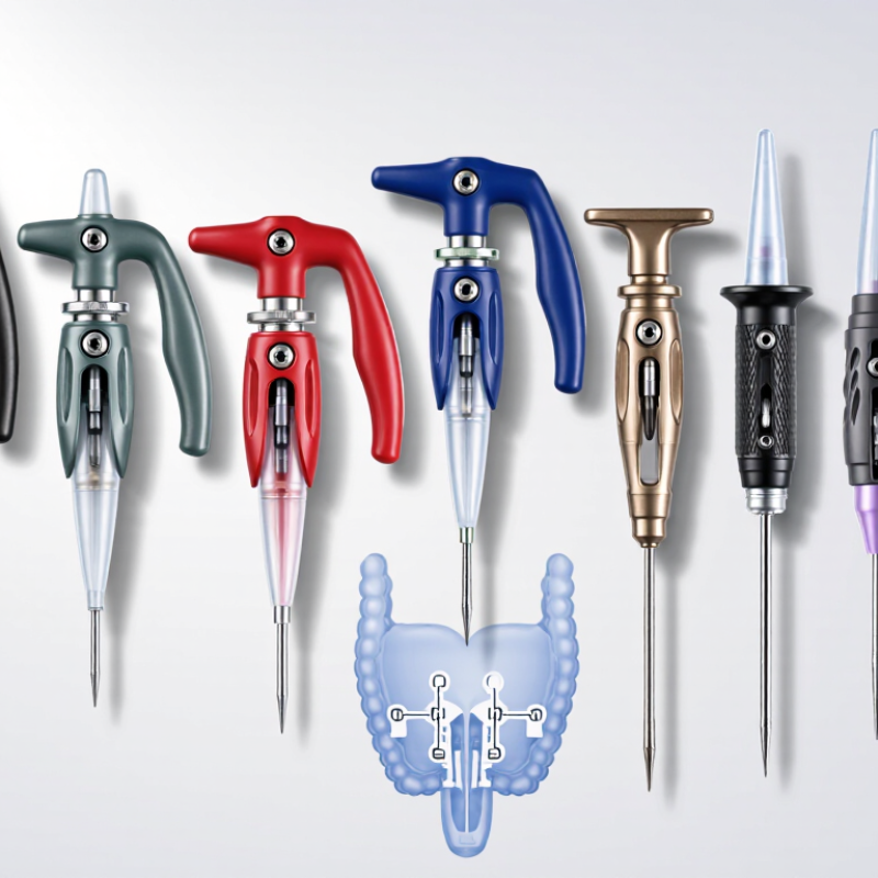
Bladeless trocars are access devices with rounded, conical tips rather than cutting blades. They separate abdominal tissue fibers instead of slicing them. This design can reduce sharp fascial disruption during laparoscopic entry. It also helps create a controlled pathway for instruments and camera systems.
Their use is practical. Surgeons may select them when they want less tissue cutting, reduced bleeding, or improved port stability. However, bladeless does not mean harmless. A Cochrane Database review found low-certainty evidence when comparing bladeless and bladed trocars. Clear superiority was not established across major complications. Published studies commonly report trocar-site hernia rates below 2%, although larger ports and poor closure can increase risk. The number matters, but technique matters more.
Insertion still requires careful angle, force control, and awareness of anatomy. A blunt tip can resist entry, then advance suddenly. That assumption is too neat. In operating-room practice, teams should inspect the port site, limit unnecessary enlargement, and close selected fascial defects. A 2023 systematic review in Surgical Endoscopy also linked port-site complications with obesity, diabetes, larger trocar diameter, and repeated instrument movement. Bladeless trocars may reduce cutting trauma, but they cannot replace surgical judgment, training, or follow-up.
| No. | Bladeless Trocar Configuration | How Access Is Created | Common Cannula Sizes | Port-Site Risk Addressed | Typical Clinical Use | Important Limitation |
|---|---|---|---|---|---|---|
| 1 | Conical blunt-tip dilating trocar | A tapered, non-cutting obturator separates and stretches tissue rather than using a blade. | 5, 8, 10, 12 and 15 mm | Reduces sharp fascial and muscle cutting; may help limit bleeding and tissue disruption. | General laparoscopic access, including abdominal procedures. | Insertion force can be higher in thick or scarred abdominal walls. |
| 2 | Expandable or radially dilating trocar | A narrow entry element passes through the abdominal wall, followed by radial dilation of the tract. | Usually 5 to 12 mm | Separates tissue planes and can reduce the amount of tissue removed at the port site. | Procedures where a controlled, smaller initial tract is preferred. | Correct alignment and controlled advancement are essential. |
| 3 | Blunt-tip optical trocar | A camera is positioned through the obturator so the operator can observe tissue layers during entry. | 5, 10 and 12 mm | Improves visual control of entry and may help identify unexpected tissue or adhesions. | Direct-vision laparoscopic entry when the surgical team uses an optical system. | Visualization does not eliminate the risk of vascular or visceral injury. |
| 4 | Balloon-sealing bladeless trocar | A blunt or dilating obturator creates access, while an inflatable balloon helps anchor and seal the cannula. | Usually 10 to 15 mm | May reduce cannula migration and gas leakage, supporting stable pneumoperitoneum. | Long procedures or cases requiring frequent instrument exchange. | Balloon inflation must be controlled to avoid excessive local pressure. |
| 5 | Fascial-retaining bladeless trocar | Retention features above and below the fascia help hold the cannula without a cutting blade. | 8, 10 and 12 mm | Helps reduce port movement, wound enlargement and repeated tissue loading. | Higher-load access sites and operations with substantial instrument manipulation. | The port may still require fascial closure according to size and surgical judgment. |
| 6 | Hasson-compatible blunt trocar | The access route is opened under direct control, and a blunt cannula is inserted through the secured incision. | 10 and 12 mm are common | Supports controlled open entry and reduces blind cutting during initial access. | Patients with previous abdominal surgery or suspected adhesions. | Requires a small incision and secure fascial fixation by the surgeon. |
| 7 | Low-profile 5 mm bladeless trocar | A small non-cutting obturator dilates a limited tract for a narrow cannula. | 5 mm | Smaller access diameter can reduce skin and fascial disruption compared with larger ports. | Diagnostic laparoscopy and procedures using slim instruments. | Limited instrument diameter and reduced suitability for specimen extraction. |
| 8 | 12 mm specimen-access bladeless trocar | A larger blunt or dilating obturator creates a working channel without a cutting blade. | 12 mm | Can provide a stable access route while avoiding additional sharp enlargement of the port. | Procedures requiring larger instruments or limited specimen removal. | Larger ports have greater potential for port-site hernia and may need fascial closure. |
| 9 | 15 mm or larger high-flow bladeless trocar | A large non-cutting dilator creates an access channel designed for high-flow suction, stapling or extraction. | 15 mm and above | The bladeless entry can limit additional sharp injury during creation of a large port. | Advanced laparoscopic procedures and larger instrument systems. | Large-diameter ports require careful closure and have higher wound-related considerations. |
| 10 | Low-friction, valve-sealing bladeless trocar | A blunt or dilating entry element is combined with a valve system that maintains the insufflation seal. | 5, 10 and 12 mm | Helps reduce gas leakage, instrument drag and unnecessary port manipulation during exchanges. | Long laparoscopic operations with repeated instrument changes. | Valve performance depends on correct instrument size, maintenance and insertion technique. |
Bladeless trocars are access devices with rounded, conical tips. They separate abdominal tissue fibers instead of cutting them. This creates a pathway for cameras and surgical instruments. Bladeless does not mean harmless.
Surgeons may choose them to reduce sharp tissue disruption and bleeding. They can also support stable instrument access. The expected benefit depends on anatomy, technique, and trocar size. Device design is only part of the outcome.
They may reduce cutting-related injury during entry. However, they cannot prevent every complication. Insertion angle, force, scars, obesity, and diabetes still matter. Risk is reduced, not removed.
Published studies commonly report rates below 2%. Larger ports and poor fascial closure can raise the risk. Port location and patient factors also influence healing. The percentage alone does not tell the whole story.
Review the patient’s anatomy, scars, body shape, and planned port locations. Use stable positioning and a suitable incision size. Advance with controlled pressure and a steady direction. Stop when unsure.
The surgeon should avoid excessive downward force. A blunt tip can resist entry, then advance suddenly. Reassess the angle, tissue resistance, and surrounding anatomy. A rushed push is not a reliable solution.
The port site should be inspected before removal. When practical, withdraw the trocar slowly under direct vision. Check for bleeding, tissue entrapment, or unexpected damage. Small details matter.
Closure may be needed for larger ports or higher-risk locations. Obesity, diabetes, and repeated instrument movement may increase concern. Follow the local surgical protocol and assess each patient individually. Technique still matters.
Bladeless trocars are access devices designed to create laparoscopic entry with a spreading or dilating tip rather than a sharp cutting blade. They are used to help reduce tissue disruption, bleeding, and fascial damage during minimally invasive procedures. Their safety advantages come from features such as tapered tips, controlled dilation, stable sleeves, secure seals, and clear depth or positioning indicators. How to reduce port-site complications with bladeless trocars? The key is to match the trocar diameter, length, tip design, and sealing system to the patient, surgical site, and procedure. A suitable device should provide reliable access while limiting unnecessary force and maintaining instrument stability.
Safe placement requires careful anatomical assessment, appropriate insertion angle, controlled advancement, and confirmation of secure positioning before instrument exchange. Surgeons should avoid excessive pressure, monitor resistance, and use visualization or established access techniques when appropriate. During removal, instruments should be withdrawn carefully, the incision inspected, and fascial closure considered according to port size and patient risk. These practices, combined with proper training and consistent monitoring, can help reduce bleeding, leakage, herniation, and other port-site concerns.

 Centuri Medical